1.3 The opponent process
The three cone signals are not what we experience. Before they leave the retina they are recombined into three new signals: one for light against dark, one for red against green, and one for blue against yellow. Each of the two color signals can swing one way or the other but not both at once. So there is no reddish green and no bluish yellow, and there are exactly four hues that look like nothing but themselves: a red that is neither orange nor purple, a yellow that is neither greenish nor orange, a green, and a blue. The rest of the brain’s work on color, in the visual cortex, builds on these three channels. The arrangement explains why yellow, which comes from the long and middle cones together, feels like an elementary color, and why the color spaces used for measuring paint are built around a red-green axis and a yellow-blue axis.
Those four hues are called the unique hues, and they can be pinned down by experiment. Hurvich and Jameson asked observers to cancel the redness, greenness, yellowness or blueness of a light by adding some of the opposite hue, which let them find where each color channel falls silent. A blue with no trace of green or red, a green that is neither yellowish nor bluish, and a yellow that is neither reddish nor greenish each turned up at a single wavelength of the spectrum, near 475, 500 and \(577\,\mathrm{nm}\) [35]. Later studies of many observers agree to within a few nanometers, with about as much spread from one person to the next [65]. No single wavelength gives a unique red: a red that is neither orange nor purple needs a little violet light added to a long-wavelength red.
The channels are drawn in Figure 3(a) for light of one wavelength at a time. Each color curve rises above zero for one hue of its pair and falls below zero for the other. The red-green channel leans to red at both ends of the spectrum and to green in the middle, so violet light looks a little reddish. The yellow-blue channel leans to yellow over the middle and long wavelengths and to blue at the short end. Where the red-green curve crosses zero, the light has no redness or greenness and looks purely yellow or purely blue; where the yellow-blue curve crosses zero, it looks purely green. The dotted lines mark these unique hues as the experiments found them. In panel (b) the same four paints as before are broken into the three channels. Cadmium yellow sends a strong yellow signal and hardly any red or green. Phthalo blue sends a blue one with little red or green, chromium oxide a dark green tinged with yellow, and quinacridone magenta, straight from the tube, a red with little yellow or blue.
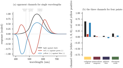
Figure 3. (a) The three opponent channels for light of one wavelength at a time: light against dark, red against green, and yellow against blue, each scaled to its largest value. The dotted lines mark the unique hues at 475, 500 and \(577\,\mathrm{nm}\), where the channels measured in experiments cross zero [35]. (b) The three channels for four paints in daylight, scaled so that a white gives full lightness and no color.
Hering proposed the opponent process in 1878, from the look of colors alone: no color ever looks both red and green, or both yellow and blue [32]. His idea long seemed to compete with the theory of Young and Helmholtz that the eye has three kinds of receptor, until Hurvich and Jameson showed in 1957 that the two fit together, with three kinds of cone at the front and opponent channels built from their signals behind [35]. Cells in the retina that compare the cones in just these ways have since been found [19]. Further on, in the visual cortex, cells compare the color of each patch with the colors around it [79], and our sense that a surface’s color belongs to the object, and stays with it when the light changes, is built from those comparisons.
The two color channels also explain why the hues form a circle. Hue runs red, yellow, green, blue and back to red through the purples, because every hue is a direction in the plane of the red-green and yellow-blue signals, and going all the way round brings you back to where you began. The purples are not in the spectrum at all. A surface looks purple only by sending both color signals toward red and blue at once, which means reflecting both ends of the spectrum and absorbing the middle. Quinacridone magenta’s curve in Figure 1(c) has that shape, but straight from the tube its violet is too weak to count and it reads as a red; with white it moves toward magenta (Section 3.3).
Yellow’s special place comes from the channels too. To the cones yellow is nothing special, a roughly equal excitation of the L and M cones with almost none of the S, but to the opponent channels it is one end of an axis, as basic as red, green or blue, and that is the level at which we name colors. Red, yellow, green and blue are the four primaries of perception. Part 4 shows that they are not the primaries of mixing paint.
Sources
[19] D. M. Dacey, Parallel pathways for spectral coding in primate retina, Annual Review of Neuroscience 23 (2000), 743–775.
[32] E. Hering, Zur Lehre vom Lichtsinne, Gerold, Vienna, 1878; English translation Outlines of a Theory of the Light Sense, L. M. Hurvich and D. Jameson, trans., Harvard University Press, 1964.
[35] L. M. Hurvich and D. Jameson, An opponent-process theory of color vision, Psychological Review 64 (1957), 384–404.
[65] B. E. Schefrin and J. S. Werner, Loci of spectral unique hues throughout the life span, Journal of the Optical Society of America A 7 (1990), 305–311.
[79] S. Zeki, J. D. G. Watson, C. J. Lueck, K. J. Friston, C. Kennard and R. S. J. Frackowiak, A direct demonstration of functional specialization in human visual cortex, Journal of Neuroscience 11 (1991), 641–649.